THE LAB AUTOMATION EXPERTS

973-376-7400

Search
Close this search box.

Protein Crystallography

Protein crystallography is used to determine the three-dimensional, atomic-level structure of proteins. It is central to biological research and structure-based drug discovery, guiding approximately 80% of lead-optimization projects, contributing to fragment screening, and supporting an increasing number of FDA submissions.

Why Automate?

As crystallography becomes more deeply integrated into drug discovery and lead optimization, teams are often required to evaluate hundreds, thousands, or even tens of thousands of conditions to meet project milestones. Automation increases throughput without adding to manual workload, while precise nanoliter dispensing enables teams to do more with limited material. Moreover, automation reduces the variability associated with manual pipetting at very small volumes, helping ensure more consistent experimental setup and results across operators. Importantly, automation does not need to be an all-or-nothing decision. It can be implemented incrementally at specific workflow steps to address bottlenecks, improve efficiency, and scale with laboratory needs.
Protein Crystallography Steps
The protein crystallography process typically involves the following steps:

1. Protein Purification

The process begins with construct design, protein expression, and purification of the recombinantly-produced protein of interest from its natural source. Techniques used during this stage may include high-throughput cloning, automated expression and purification systems, and chromatography to produce a sufficiently concentrated and pure protein sample.

2. Plating of Screens

During this step, a broad range of screening solutions is prepared and aliquoted for subsequent crystallization experiments. Automated liquid handlers improve accuracy and reproducibility during this process while enabling efficient preparation at scale.

Scorpion™ Screen Builder:
Scorpion™ Screen Builder is a high-speed single-channel automated pipetting robot designed for crystallization screen preparation and optimization. Its dedicated software supports reagent transfer from varied labware into SBS-format plates and enables customized matrices across pH, precipitant concentrations, additives, and reagent combinations.
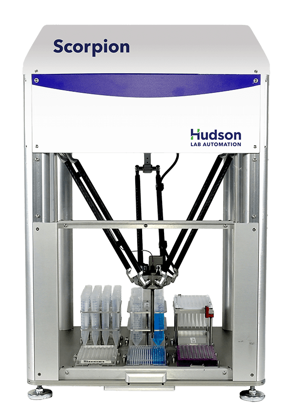
Hudson Scorpion Single-Channel Pipettor

3. Protein Drop Setup

Purified protein is dispensed and mixed with selected screening solutions in crystallization plates. There are several experimental formats available for producing diffraction-quality crystals.
  • Sitting Drop Vapor Diffusion: A small protein-reagent drop is placed on a pedestal in a sealed well above a reservoir containing a higher concentration of precipitant. As the system equilibrates, water vapor gradually leaves the drop, increasing the protein and precipitant concentrations until crystals form.
  • Hanging Drop Vapor Diffusion: A small protein-reagent drop is suspended from an inverted coverslip above a reservoir containing a higher concentration of precipitant. Vapor diffusion gradually concentrates the drop, creating the supersaturation required for protein crystals to nucleate and grow.
  • Microbatch: Protein and precipitant are mixed in tiny droplets under oil. This limits evaporation and provides a stable, sealed environment for nucleation and crystal growth that minimizes the concentration changes associated with vapor diffusion.
  • Seeding: Crystals or crystal fragments are introduced into a fresh, supersaturated drop to control nucleation and promote growth of larger, better-ordered crystals.
  • Lipidic Cubic Phase (LCP) in meso Crystallization: Membrane proteins are reconstituted within a viscous LCP composed of a continuous lipid bilayer that mimics the cell membrane. This environment helps stabilize membrane proteins in their native conformations while providing a suitable matrix for crystallization.
  • Sponge Phase: A more easily handled liquid analog of the LCP that allows crystallization of membrane proteins with larger hydrophilic domains.
  • Bicelle: Membrane proteins are incorporated into small, disc-shaped lipid bilayers (bicelles) that maintain a native-like environment and facilitate crystallization.

Crystal Gryphon™:
The Crystal Gryphon™ is a protein crystallization pipettor that can be easily adapted to perform all of the assays listed above. It provides rapid 96-well plate setup, precise nanoliter dispensing, flexible reservoir and optimization workflows, and reproducible performance without using disposable tips.
This product was formerly known as the ARI Crystal Gryphon.
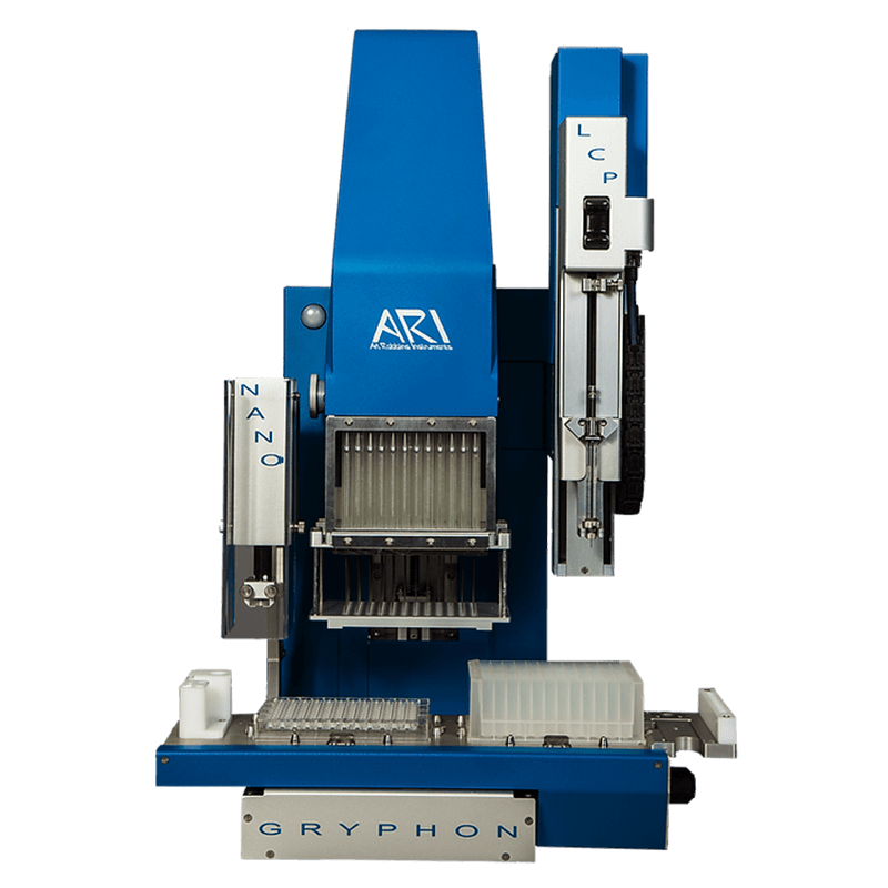
ARI Crystal Gryphon Protein crystallization pipettor

INTELLI-PLATE® Protein Crystallization Plate:
The INTELLI-PLATE® is designed for automated vapor-diffusion crystallography and pairs directly with the Crystal Gryphon™. It is available in 96-, 48-, and 24-well formats and offers high optical clarity, reduced evaporation, and flexible one-, two-, or three-drop configurations.
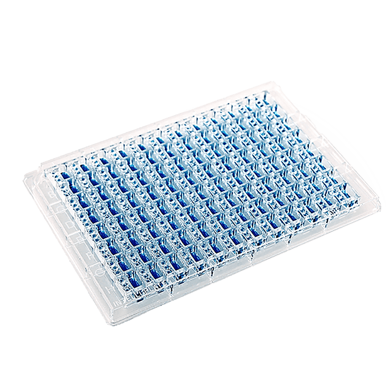
ARI intelli-plate crystallization plate.

4. Incubation

Plates are incubated under controlled conditions to allow the protein and screening solutions to equilibrate. During this period, protein molecules nucleate and grow into crystals that can be evaluated for diffraction.

PlateCrane™ SciClops™ Robotic Arm:
The PlateCrane™ SciClops™ Robotic Arm enables continuous, hands-free plate transfers between incubators and imaging systems, reducing repetitive, time-consuming handling during crystallization workflows.
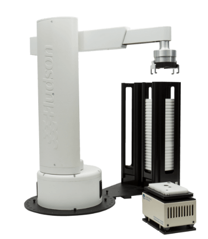
PlateCrane SciClops Higher Capacity Robotic Arm

5. Imaging

Plates are viewed under visible and ultraviolet light to monitor crystal growth over time, detect microcrystals, and distinguish protein crystals from salt crystals. Automated imagers allow researchers to compare development between imaging sessions, maintain digital experiment records, and quickly identify conditions suitable for further optimization.

CrysCam UV Imaging System:
The CrysCam UV Imaging System is an automated crystal imaging platform that captures aligned visible-light and ultraviolet images across multiple focal planes and enables users to flag wells of interest. With the CrysCam, researchers can use any SBS-format plate as well as Linbro plates and benefit from integration with the PlateCrane™ SciClops™ Robotic Arm.
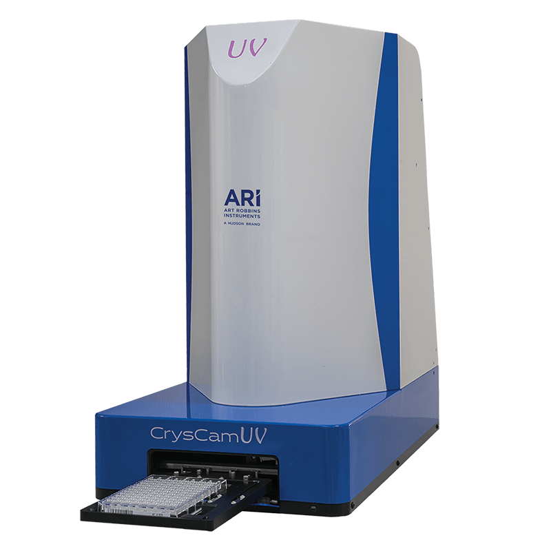
ARI CrysCam UV Imaging System

6. Optimization

Crystal optimization refines promising crystallization conditions to improve crystal size and diffraction quality. Optimization can improve crystal packing, a key determinant of data quality, and typically involves preparing large numbers of customized matrices that vary in parameters such as pH, precipitant concentration, additives, and reagent combinations. The Scorpion™ Screen Builder automates customized optimization screens, reducing manual effort while improving reproducibility and efficiency.

7. X-ray Diffraction

Protein crystals are harvested, mounted, and subjected to X-ray diffraction analysis. X-rays are directed at the protein crystals, and as they pass through the crystal lattice, they scatter in different directions. The resulting diffraction pattern contains information about the arrangement of atoms within the crystal. Diffraction data are often collected at synchrotron facilities, which provide intense, highly focused X-ray beams.

8. Data Collection

The diffraction pattern is captured using a detector, such as a charge-coupled device (CCD) camera or hybrid pixel-array detectors (HPADs). Multiple diffraction images are collected from different orientations of the crystal, providing overlapping data.

9. Structure Determination

The collected diffraction data provide the intensities of X-rays diffracted by the protein crystal, from which the magnitudes of the structure factors (amplitudes) can be derived. However, the corresponding phase information is not measured directly. Phase information can be obtained using methods such as molecular replacement (MR), which uses a structurally related protein model to estimate the phases. Structure determination may involve experimental phasing methods such as single-wavelength anomalous diffraction (SAD) and multi-wavelength anomalous diffraction (MAD), which exploit anomalous scattering from specific atoms in the crystal. The measured diffraction amplitudes and estimated phases are then combined to calculate an electron density map, which represents the spatial distribution of electron density within the crystal. This map is used to build an atomic model of the protein, assigning the positions of atoms to regions of electron density.

10. Model Building and Refinement

Atomic models of the protein are built based on the electron density map. Initially, a rough model is constructed, and then it is refined iteratively to fit the experimental data more accurately. This refinement process involves adjusting the positions of atoms to minimize discrepancies between the observed and model-calculated electron densities. Predicted models from tools such as AlphaFold may support model building or provide starting structures for molecular replacement.

11. Validation

The final atomic model is validated using various criteria to ensure its accuracy and reliability. This validation process may include assessing the quality of the electron density map, checking for stereochemical correctness, and comparing the model with other experimental data if available. It may also include reviewing B-factors, which indicate the relative mobility or positional uncertainty of atoms in the model.

12. Publication and Further Analysis

Once validated, the final crystal structure of the protein is typically submitted to the Protein Data Bank for use by the broader scientific community and published in scientific journals, making it available to the scientific community for further analysis and interpretation. The structural information obtained from protein crystallography can provide insights into the protein’s function, mechanism of action, and potential applications in drug discovery and biotechnology.
The Future of Structural Biology

Structural biology will see significant improvements in efficiency as AI and automation enable faster decision-making, greater workflow integration, and more scalable crystallography processes.

AI is helping improve crystallography efficiency by learning from screening and optimization results and using that data to recommend new conditions and guide subsequent experiments. This enables researchers to make faster, data-driven optimization decisions and identify promising conditions more efficiently.

Automation facilitates greater integration of individual processes across the entire protein crystallography workflow. This minimizes the manual handling steps normally required between different processes, leading to reduced manual burden for laboratory personnel, and fewer sources of variability which may lead to confounding results and misguided decision-making. The future points toward more fully connected, end-to-end workflows spanning robotics, imaging systems, beamlines, and data-analysis platforms. Today, teams can automate specific workflow steps based on their needs and gradually build toward fully connected, end-to-end protein crystallography.

What used to take 45 minutes...

now takes 25. Automatically.

Scorpion™

Single-Channel Automated Pipetting Robot

Get the details in "Adaptable Automation for Rapidly Changing Labs"

Hudson Scorpion Single-Channel Pipettor