This blog highlights the key points covered in an Xtalks webinar featuring Matt Cucuzza, Marketing Manager at Hudson Lab Automation, and Meagan Dufrisne, Structural Biologist at Helix BioStructures. Their discussion explored the enduring value of protein crystallography across life sciences applications and how automation improves accuracy, throughput, and scalability throughout the workflow.
X-ray crystallography generates high-resolution electron-density maps that support accurate protein structure determination and ligand placement. It is a fundamental technique in various life sciences and biotechnology applications. A principal example is drug discovery, where it remains a workhorse for high-throughput protein-ligand structure determination, guiding 80% of lead optimization projects and supporting a growing number of FDA submissions. As cryo-electron microscopy expands the range of biological targets that can be studied, X-ray crystallography remains essential for obtaining high-resolution structural data for lead optimization, fragment screening, and biologics development, among other applications. Rather than replacing X-ray crystallography, the growth of cryo-electron microscopy is increasing the need for crystallography as more targets enter structural biology workflows.
Its ongoing significance is reflected in the rise in worldwide X-ray crystallography structure submissions to the Protein Data Bank, from 9,566 in 2015 to 10,865 in 2025. Protein crystallography’s role in drug development is set to grow, with a projected market expansion from USD 1.8 billion in 2025 to USD 3.0 billion by 2032. This growing demand is increasing the need for process optimization to improve accuracy, reduce manual workload, and support scalable operations. Automation has emerged as a key approach to addressing these workflow challenges and represents a natural evolution in protein crystallography as teams seek to increase throughput while improving accuracy and reproducibility.
In the early 2000s, crystallography workflows were largely manual, limiting throughput to around 100 pipetted conditions per day. Automated liquid handlers introduced in 2003 substantially increased capacity, while automated imaging in 2007 reduced the need for time-consuming microscope inspection. Remote-access and mail-in synchrotron services followed in 2011, broadening access to high-quality X-ray diffraction, and AlphaFold transformed structure prediction and model building in 2020. Automated data-processing pipelines also accelerated molecular replacement, model building, and structural refinement. By 2026, these advances are converging into integrated workflows spanning sample preparation, crystallization, imaging, data collection, and AI-assisted crystallography.
Statistics that illustrate the progress over the last quarter-century include:
Key business benefits of these improvements include:
Protein crystallography is a complex, multistep process in which inefficiencies or bottlenecks at any stage can delay downstream activities and extend development timelines. This section outlines the key workflow steps and highlights where bottlenecks can arise.
This step produces the high-purity protein required for crystallization and downstream analysis. However, certain targets, including membrane proteins, can be difficult to isolate in sufficient quantities and suitable conformations. Bottlenecks can emerge when low yields, instability, or lengthy purification steps delay crystallization or limit the number of conditions that can be tested.
This step involves combining purified protein with screening solutions to form crystallization drops and testing a broad range of conditions to identify those that produce diffraction-quality crystals. Throughput can become a major bottleneck because hundreds or thousands of conditions may require screening and pre-use aliquoting.
This step involves inspecting each drop to identify crystal formation and assess crystal quality and suitability for diffraction. Manual inspection can limit throughput, while repeatedly transferring large numbers of plates between incubators and imaging systems can create additional delays.
This step involves refining crystallization conditions to improve crystal size and diffraction quality. Historically, throughput was constrained by 24-well trays, larger drops requiring substantially more protein, and the manual preparation of custom optimization solutions.
Automation has significantly improved the accuracy, throughput, and scalability of protein crystallography. However, deciding when to implement automation, and which stage of the workflow to target, remains a complex decision shaped by time requirements, reproducibility, and future capacity. A fully automated workflow can free up researcher time and support greater scale, but instrumentation also brings substantial upfront and ongoing operational costs. This can make it difficult to identify the point at which automation becomes the most strategic investment. It is also important to consider the opportunities automation enables, including testing more conditions, taking on additional projects, expanding operations, and pursuing more ambitious funding opportunities. Ultimately, while investment timing and specific needs will vary by laboratory, consistent reproducibility, time gains, and real scalability can only be achieved through automation.
Automated protein crystallography workflows allow teams to progress more quickly with large-scale screening but also provide advantages for more challenging projects as the following real-world case studies demonstrate.
Using manual methods, setting up more than 45,000 crystallization drops would require nearly eight months of laboratory work. By leveraging automation, Helix was able to screen more than 45,000 drops and deliver 11 structures in just 11 weeks, ultimately completing 20 structures for the project. This demonstrates how automation can make large-scale crystallography projects more feasible while substantially reducing timelines, with downstream benefits for the cost and efficiency of drug development.
Low-yielding and conformationally unstable proteins have long been a major challenge in protein crystallography workflows. Helix worked with a particularly challenging kinase that required ligands to maintain solubility and had to be purified within a single day using multiple chromatography steps, only to yield 1.5 mg from a 20 L culture. Despite the limited protein available, Helix generated four plates comprising 384 conditions and 1,152 drops, ultimately producing a crystal structure with a ligand of interest bound. Achieving this combination of speed and experimental scale would have been extremely difficult with manual methods. This real-world example highlights how automation enables discovery in addition to increasing throughput.
Hudson Lab Automation provides solutions that address specific workflow bottlenecks, improve efficiency and scalability, as well as supporting complete end-to-end automation. Below are the key points where our instruments can integrate into the crystallization workflow.
Traditional liquid handlers are designed for biological workflows—not chemistry.
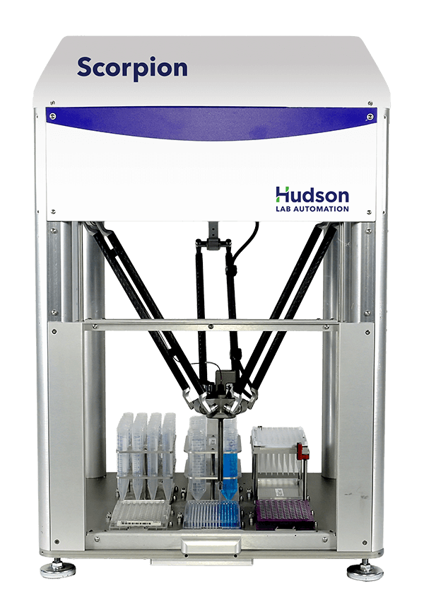
Scorpion™ Screen Builder: Automates crystallization screen preparation and optimization, transferring reagents from varied labware into SBS-format plates and generating customized matrices across pH, precipitant concentrations, additives, and reagent combinations.
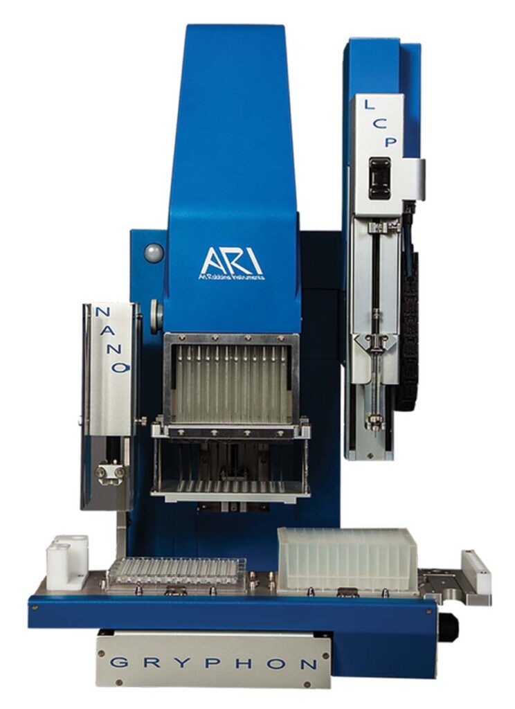
Crystal Gryphon™: Automates crystallization drop setup, preparing 96-well plates in under a minute while dispensing nanoliter protein volumes precisely, reproducibly, and without using disposable pipette tips.
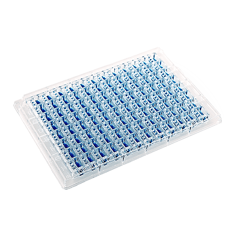
Supports automated vapor-diffusion crystallography with optically clear, low-evaporation plates in 96-, 48-, and 24-well formats, including flexible one-, two-, or three-drop configurations for varied experimental designs.
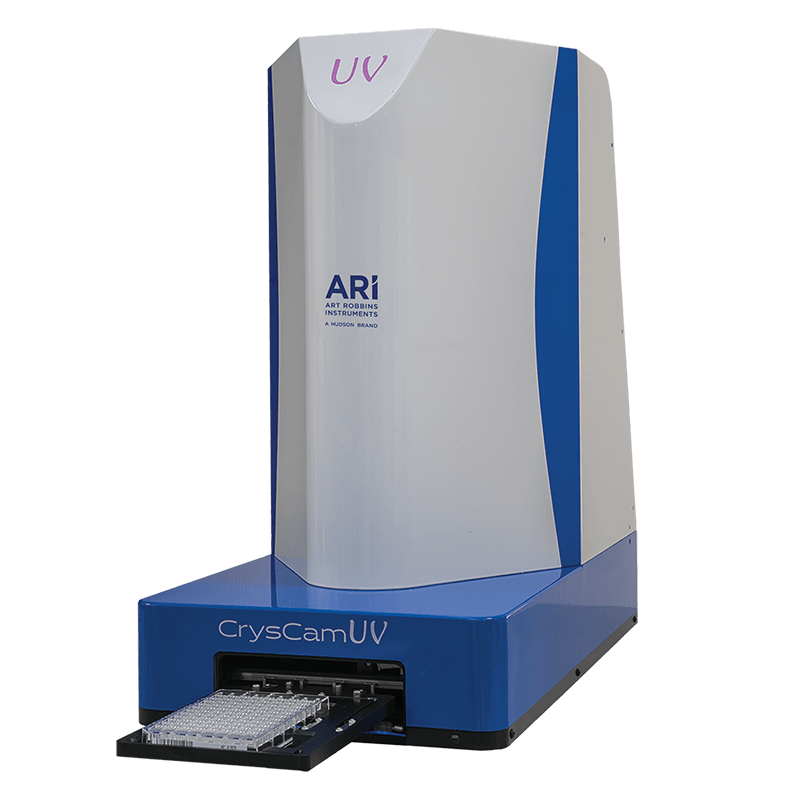
CrysCam UV Imaging System: Automates crystal imaging with visible and ultraviolet views, multiple focal planes, and broad plate compatibility.
Copyright 2026 ® Hudson Lab Automation
Get the details in "Adaptable Automation for Rapidly Changing Labs"